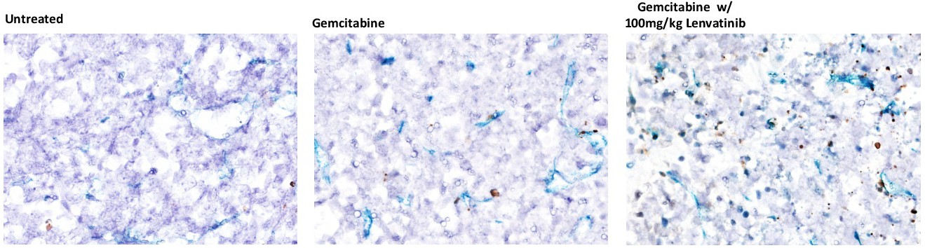
Fig. 4. Lenvatinib increases endothelial cell apoptosis after gemcitabine in vivo. Lenvatinib was administered to sv129/BL6 mice harboring 100mm3 MCA/129 fibrosarcoma flank tumors and 1h later mice were treated with 240 mg/kg of gemcitabine. After 4h mice were sacrificed, and 5-μm thick tumor sections were double-stained using TUNEL labeling and MECA-32 Ab to identify apoptotic endothelial cells (ECs). Representative fields are shown. Apoptotic endothelial cells exhibit a brown TUNEL-positive nuclear signal surrounded by a dark blue plasma membrane signal for MECA-32.